
Ultrasound: How it can help your pet
The vet’s office
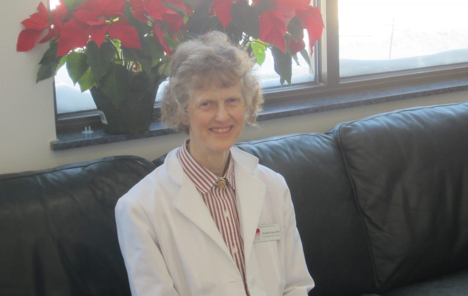
By Dr. Elizabeth Knabe, DVM
Wildwood Animal Hospital and Clinic LLC
Beginning about 30-40 years ago, ultrasound technology started coming into practice in veterinary referral centers and teaching hospitals. Since then it has also become a diagnostic tool offered by private practices and emergency clinics.
A special probe on a unit gives off ultra-high-frequency sound waves that are sent into the body. The probe then “listens’’ for the echoes of the sound waves returning and interprets them as an image of what is inside the body. Just like with a submarine using sonar, the echoes coming back tell what the structures are like and how far apart they are. The image is formed in black, white, and shades of grey on a computer monitor. The images can be stored as computer files and viewed again or sent to specialists for additional consultations.
Ultrasound can give details of what lies within organs. A veterinarian may suspect something is wrong with a pet’s internal organs based on an exam and blood tests. Whereas an abdominal X-ray will show the outline of the organ, an ultrasound can often show if there are lumps or cysts within the organ. An additional step may be a special biopsy with a thin needle directed into the lump. The ultrasound will help guide the needle to the site. A pet may then be able to get a diagnosis without having to undergo the stress of surgery.
Ultrasound also has an advantage over MRI and CT scans in that absolute stillness is not required. People can lie still for the time to run a MRI or CT, but pets cannot and need general anesthesia or heavy sedation. For the ultrasound, some pets will be given a mild sedation, but many that are calm to begin with allow an abdominal scan while fully awake. If a pet moves during an ultrasound scan, the operator just waits for the pet to return to lying quietly.
Fur over the abdomen will need to be clipped for an ultrasound, however. The sound waves are scattered and useless if they try to go through hair. Once the hair is clipped, a special gel is placed on the probe and pet’s skin, which allows the maximum transmission of sound waves into the body.
Besides the abdomen, ultrasound is used to study the heart. There are many heart conditions where a heart echo study is the main way to diagnose it so that appropriate therapies can be started.
Ultrasound does not completely replace radiographs though. X-rays are still used to study bones, and chest X-rays show details in the lungs.
Increasingly, ultrasound is becoming a useful diagnostic tool to reach for earlier rather than later.
Wildwood Animal Hospital and Clinic LLC is located at 210 Airpark Road in Marshfield and online at wildwoodanimalhospital.net.

Leave a reply
You must be logged in to post a comment.